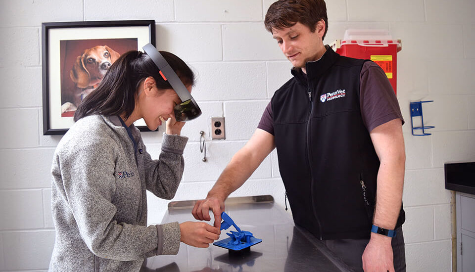
Beginning this spring, Penn Vet is launching an augmented reality interface that will help students learn how to perform challenging spinal cord surgery, writes Sacha Adorno for Penn Vet News.
The program is the newest addition to Penn Vet’s growing portfolio of advanced technology for teaching and clinical care.
The idea for the program came from the school’s staff neurologist, Dr. Jonathan Wood. At the time, he and Dr. Evelyn Galban, Clinical Assistant Professor of Neurology and Neurosurgery, were treating a young dog with a large tumor on her skull.
“I wanted to understand the tumor’s position and how to proceed before actually operating,” said Dr. Wood.
[uam_ad id=”71803″]
Advertisement
With the growing library of 3D models, Wood decided to take it a step further. He became interested in augmented reality, which is the interplay between animated and real objects.
He approached Dr. Stephen Lane, Director of the university’s Computer Graphics and Game Technology Masters Program. They have now developed an augmented reality program that animates a canine spinal cord for a procedure called atlantoaxial stabilization.
This program will allow students to practice on an animated spinal cord until they master the procedure.
Read more about augmented reality at PennVet News here.
[uam_ad id=”72786″]


")


")


")


")